
Planning
We go through the results with you on screen. Before treatment starts, you receive a written treatment and cost plan (Heil- und Kostenplan).

All five steps take place at the practice in Schüttaustraße. Dr. Markiel Haimov is a dentist and a dental technician: he plans from the X-ray and knows how the crown is made.
A cone beam CT scan (CBCT, in German: DVT) shows the jawbone, the course of the nerves and the tooth roots in three dimensions. We use it to measure the width, height and depth of the bone.
The scan takes about 20 seconds. The machine is open-sided, and only the head area is X-rayed. The radiation dose is lower than with a CT scan.
We use it before implants, when root fractures or cysts are suspected, and when a normal X-ray is not enough. You do not need an appointment with a radiologist.
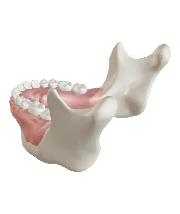
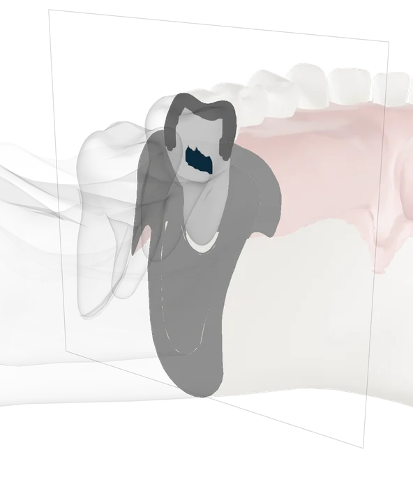
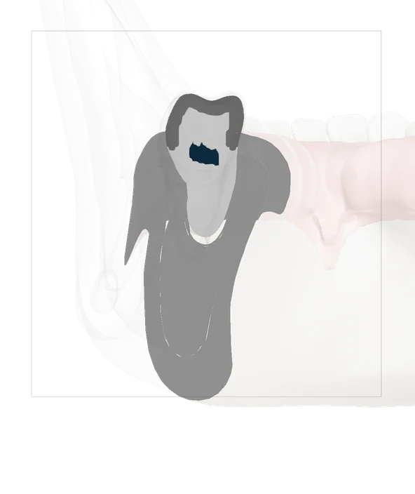
We go through the results with you on screen. Before treatment starts, you receive a written treatment and cost plan (Heil- und Kostenplan).

An intraoral scanner moves a camera along the upper and lower jaw without touching the teeth. It records the teeth digitally, without impression material. The scan takes about ten minutes.
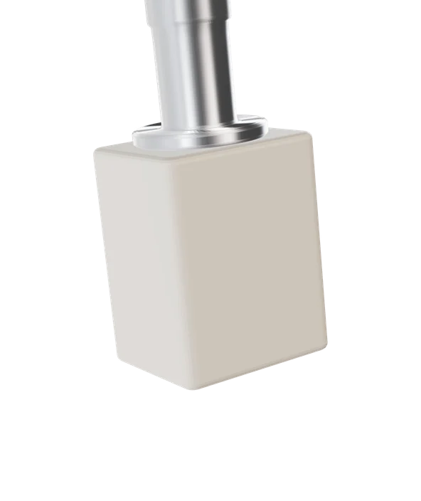
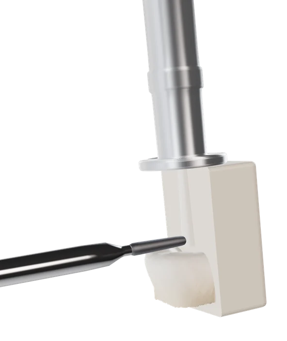
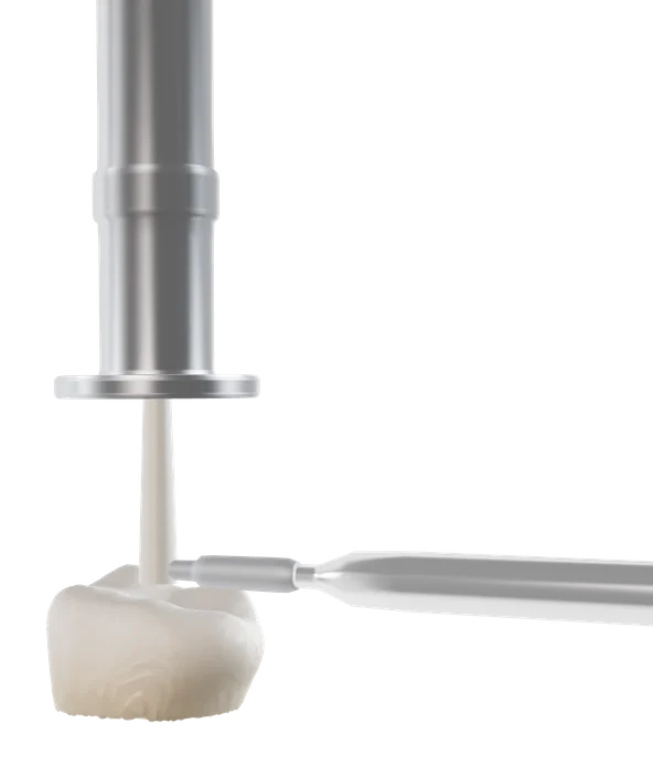
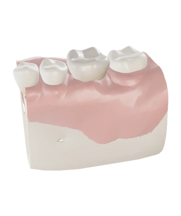
From the scan, the crown is designed on the computer and milled from a ceramic block at the practice. The system is called CEREC.
This produces ceramic inlays, partial crowns, crowns and bridges in one session, without a temporary and without a second anaesthetic. Milling takes about 20 minutes, the whole treatment about 90 to 120 minutes.
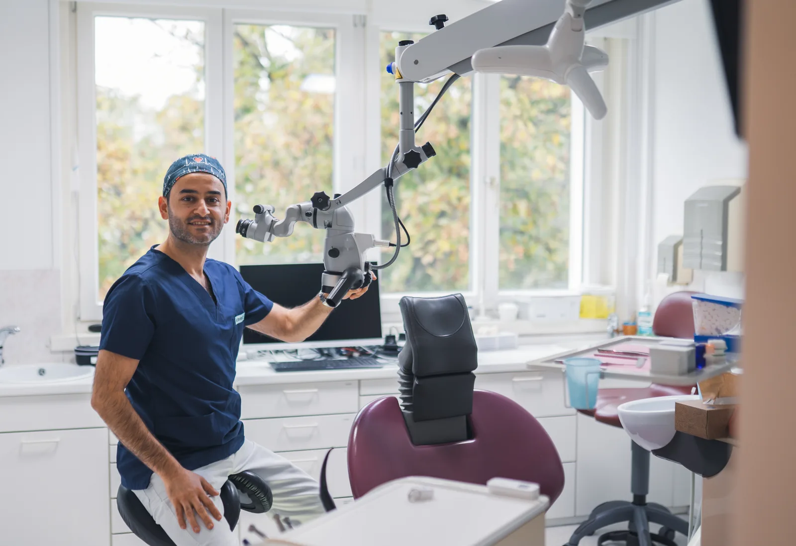
The dental microscope magnifies up to 25 times and lights the tooth without shadows. We use it mainly for root canal treatment, to find the fine canals too.
A camera on the microscope records what happens inside the tooth.
Before treatment starts, the treatment and cost plan sets out what using this equipment costs and what your insurer covers.
A cone beam CT scan, a 3D X-ray of the jaw, teeth and nerves. It shows structures that overlap on a normal X-ray.
Usually not; it is mostly a private treatment. We only do it when your treatment needs it.
Scan, design, milling and fitting at the same appointment. You need no temporary and no second appointment.